Creative Minds 𖹭 Happy Hearts

Plant Cell Plasmolysis Lab Sheet, Red Onion Microscope Experiment
Red onion cells visibly shrink away from their walls in a salt solution, and this plant cell plasmolysis lab sheet gives your student a structured way to capture exactly what they see.
Designed for grades 6-9, this plant cell plasmolysis lab sheet walks your homeschooler through a complete microscopy experiment using red or purple onion epidermis. Students form a hypothesis, prepare a slide in distilled water, observe under low and high power, then introduce a 10% salt solution to trigger plasmolysis and draw both the before and after cell states. A labeled reference diagram shows what the cell wall, membrane, vacuole, and cytoplasm should look like at each stage, so students know what they are actually searching for under the lens.
The results table asks students to compare cell membrane position, vacuole size, cytoplasm appearance, and whether plasmolysis was observed, making this plant cell plasmolysis lab sheet a strong fit for a unit study on osmosis, cell structure, or plant biology. Two Think and Extend questions push beyond the observation, asking why water moved out of the cell and where plasmolysis matters in real-world plant health. A conclusion prompt ties findings back to the original hypothesis, giving your student practice in scientific reasoning from start to finish.
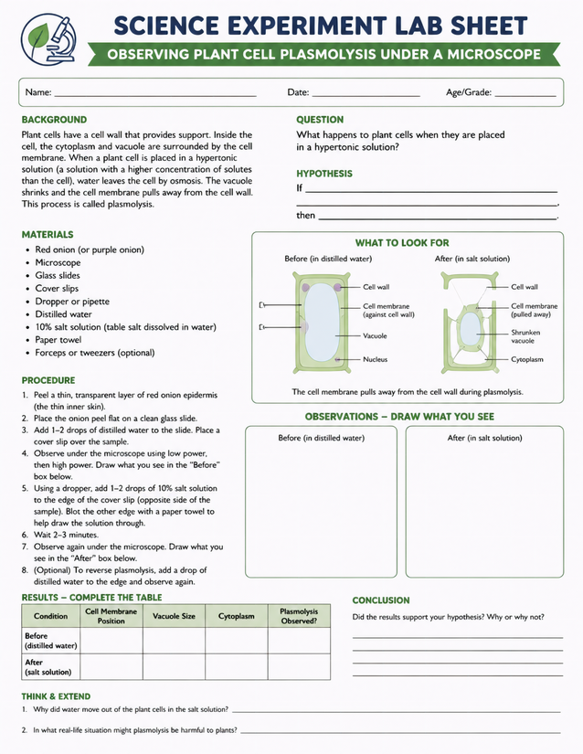
Record before-and-after microscope observations with this plant cell plasmolysis lab sheet, designed for grades 6-9 homeschoolers studying osmosis.
Exploring the Plant Cell Plasmolysis Lab Sheet with Red Onion

Usage Notes
Nearly everything on the materials list is already in a typical household kitchen: red or purple onion, table salt dissolved in water, paper towel, and forceps or tweezers. The specialty items are glass slides, cover slips, and a compound microscope capable of low and high power. The procedure involves adding salt solution to a live slide sample, so remind students to handle glass slides carefully and work over a stable surface.
Printing Tips
Standard 20 lb copy paper works for the observation drawing boxes, but 24 lb paper takes pencil and pen more cleanly if students are drawing detailed cell sketches. Slip the sheet into a clipboard for microscope-side note-taking, or insert it into a dedicated lab notebook after the session is complete.