Creative Minds 𖹭 Happy Hearts

Making a Model of Mitosis with Clay, Five Stages
Building all five stages of cell division with your hands makes mitosis stick in a way a diagram simply cannot. Making a model of mitosis with clay gives your student a tactile sequence they can arrange, label, and explain.
Cell division can feel abstract on paper, but this lab sheet walks your student through constructing a physical clay model for each of the five mitosis stages: Interphase, Prophase, Metaphase, Anaphase, and Telophase with Cytokinesis. A clay color guide assigns specific colors to the nucleus, chromosomes, centrosomes, spindle fibers, and cell membrane, so your student builds with accuracy rather than guessing. The recording chart asks them to describe what each stage looks like in their model, what is happening biologically, and which key parts are shown. Aimed at grades 6-8, making a model of mitosis with clay fits naturally into a cells and heredity unit study and pairs well with a lesson on DNA replication before it. Four analysis questions close the sheet, asking students to reason through why cells copy DNA first, what spindle fibers do, and how mitosis benefits living things, moving them from modeling to genuine biological understanding.
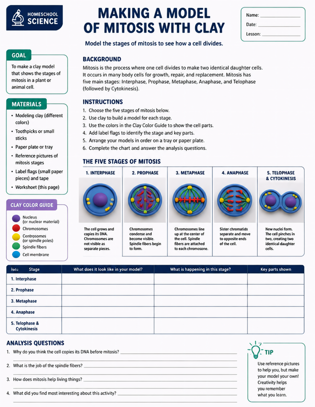
Grades 6-8 homeschoolers build all five stages when making a model of mitosis with clay. Instant download included.
Exploring Making a Model of Mitosis with Clay

Usage Notes
Multicolored modeling clay is the primary material, and the sheet specifies at least five distinct colors to match the clay color guide. Toothpicks or small sticks stand in for spindle fibers, and label flags made from small paper pieces and tape identify each stage. A paper plate or tray keeps the five finished models organized in sequence. No heat or chemicals are involved, so this is safe for independent student work without close adult supervision.

Printing Tips
A single sheet on 24 lb copy paper holds up well enough for classroom use, but 32 lb paper handles pencil notes in the recording chart without feathering when students press firmly. Slide it into a page protector inside a science lab notebook after the clay models are complete and photographed for a tidy unit record.