Creative Minds 𖹭 Happy Hearts

Observing Blood Cells Under a Microscope, Prepared Slides
Stained slides reveal three distinct cell types, each with a different shape and job. Observing blood cells under a microscope gives your student a front-row look at how the circulatory system actually works at the cellular level.
This lab sheet walks students through observing blood cells under a microscope using a prepared human blood smear slide, starting at 4x magnification and stepping up through 10x and 40x objectives. Students record shape, color, and relative size for red blood cells, white blood cells, and platelets in a structured observation table, then draw and label each cell type inside a provided circle. The sheet suits grades 5-8 and aligns naturally with a human body or life science unit study.
The background section gives students enough context to know what they are looking for before they touch the microscope, and the eight-step procedure keeps the session orderly without hand-holding. Two conclusion questions ask students to reflect on what they learned and how the three cell types cooperate to maintain health, making observing blood cells under a microscope a complete standalone lesson rather than just a coloring exercise.
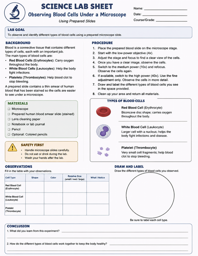
Grades 5-8 homeschoolers practice observing blood cells under a microscope using prepared slides, recording shape, color, and relative size.
Getting Started with Observing Blood Cells Under a Microscope

Usage Notes
Prepared stained blood smear slides are a specialty supply available from science education vendors, so order those ahead of time. The remaining materials are household or standard homeschool items: lens cleaning paper, a notebook or lab journal, a pencil, and optional colored pencils for the draw-and-label section. The sheet includes a Safety First box reminding students to handle slides carefully, avoid eating or drinking during the lab, and wash hands afterward.
Printing Tips
Standard 20-lb copy paper works for the observation table, but 24-lb paper holds up better if students rest their hand on the sheet while writing at the microscope stage. Print one copy per student as a loose sheet on a clipboard, then transfer it to a lab journal or three-ring binder after the session.