Creative Minds 𖹭 Happy Hearts

Observing Mitosis in Onion Root Tip Cells, Stages Chart Included
Onion roots sitting in a jar of water are the starting point for observing mitosis in onion root tip cells, one of the most visually satisfying labs in middle school biology.
Your student will grow onion roots, prepare a microscope slide using iodine or aceto-orcein stain, and then search for cells in five distinct stages of mitosis: interphase, prophase, metaphase, anaphase, and telophase. The sheet walks through an eight-step procedure and includes a labeled diagram of each stage so your grades 7-10 homeschooler knows exactly what to look for at high magnification before touching the microscope. Observing mitosis in onion root tip cells fits naturally into a cell biology unit study as the hands-on follow-up to lessons on the cell cycle, and it works equally well as a standalone lab. Completing the observation table, which asks students to count and compare cells in each stage, builds real data-recording practice, while the four analysis questions push students to explain why most cells appear in interphase and why mitosis matters to living organisms.
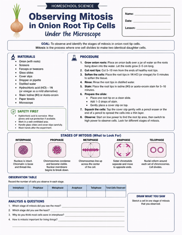
Grades 7-10 homeschoolers study cell division by observing mitosis in onion root tip cells under the microscope. Instant download.
Getting Started with Observing Mitosis in Onion Root Tip Cells

Usage Notes
Hydrochloric acid at 1N is listed on the materials section, and the safety panel flags it as corrosive, so gloves and eye protection are required and a well-ventilated workspace is non-negotiable. The sheet offers vinegar as a milder alternative, which keeps this accessible for families without lab-grade chemicals. Glass slides, cover slips, a dropper, and iodine or aceto-orcein stain are specialty supplies, but most are inexpensive and reusable across future microscopy labs.

Printing Tips
Standard 20 lb copy paper works for the observation table and analysis questions, but 24 lb or heavier holds up better if the sheet will sit near wet slides during the lab. Print as a loose sheet to clip onto a lab notebook or slide inside a page protector for reference while your student works at the microscope.